Thursday, June 28, 2007
Wednesday, June 27, 2007
Tuesday, June 26, 2007
ჩავუხტეთ ბარათაშვილსა
ი არსენა რა ყოფილა
რა არი ამისთანაო
რას იკვეხ, ბარათაშვილო,
შენი ლამაზი ქალითა,
ჩაგიმწყვდევია ცხრაკლიტულს
მზეს არ ანახვებ თვალითა.
ტალად ვეფხვები დაგისვამს
უალმასესი ბრჭყალითა,
ვერ დაიფარვენ, დაგავსებ
ქალს წაგგვრი მაჯაგანითა.
აბჯარ აისხა, შაჰკაზმო,
შენი ქურანი მალია,
ღვთის რისხვა გამამადევნო
ქვას ფრეწდის ქორის თვალითა,
ცხრა ვაჟი შამამისივო
ცხრა ალესილი ხრმალია,
ცხრა რძალი ბანზე დაგისვა
დაღრეჯით მატირალია.
შევარდნის მართვე გახლავარ
მოყმე ვარ ალვისტანაო,
ცხრა მუხა აკვანს მირწევდა
ცხრა წყარომ პირი მბანაო,
ცხრა დევი ხელით შავიპყარ
შავკოჭე, ჩავეც დანაო
ცხრა სიკვდილ ისე დავლახენ
წარბიც არ შემაქანაო.
ჩემი აკვანი კუბოა,
ზარი გლოვისა - ნანაო,
სული გვამს როცა გასცდება
მიმღერე სულთათანაო.
მერაბ კოსტავა
Monday, June 25, 2007
"O Captain" by Walt Whitman
O Captain! my Captain! our fearful trip is done;
The ship has weathered every rack, the prize we sought is won;
The port is near, the bells I hear, the people all exulting,
While follow eyes the steady keel, the vessel grim and daring.
But O heart! heart! heart!
O the bleeding drops of red!
Where on the deck my Captain lies,
Fallen cold and dead.
O captain! my Captain! rise up and hear the bells;
Rise up! For you the flag is flung, for you the bugle trills:
For you bouquets and ribboned wreaths, for you the shores a-crowding:
For you they call, the swaying mass, their eager faces turning.
Here Captain! dear father!
This arm beneath your head;
It is some dream that on the deck,
You've fallen cold and dead.
My Captain does not answer, his lips are pale and still;
My father does not feel my arm, he has no pulse nor will;
The ship is anchor'd safe and sound, its voyage closed and done;
From fearful trip the victor ship comes in with object won!
Exult, O shores, and ring, O bells!
But I with mournful tread,
Walk the deck my Captain lies,
Fallen cold and dead.
Sunday, June 24, 2007
Imaging and radiotherapy: getting closer all the time
Over the past 30 years, radiation oncologists and medical physicists have made remarkable progress with respect to both the accuracy of tumour irradiation and minimizing the levels of collateral damage to healthy tissue. This is a story of relentless innovation, with plenty of game-changing technologies along the way. The early application of computers in treatment planning, for example, was followed by the introduction of 3D imaging with CT and MRI, an advance that yielded improved delineation of target volumes, while specialized computer algorithms allowed physicists to precalculate radiotherapy dose distributions with increasing speed and accuracy.
Subsequently, linacs were equipped with computer-controllable multileaf collimators (MLCs), a core enabling technology in 3D conformal radiation therapy (CRT) and intensity-modulated radiation therapy (IMRT). Today, with 3D CRT and IMRT, it is possible to produce a highly conformal dose distribution tailored to nearly every kind of target volume, no matter how complex. In this sense, charged-particle therapy looks like providing the next big step forward, with protons "tuned" to deliver most of their energy at a specified depth, thereby reducing the chances of skin burns and damage to healthy tissue surrounding a tumour.
Yet despite all this technological and clinical innovation, experience shows that in many cases it is still not possible to control local tumour growth. There's a simple explanation for this anomaly, summed up neatly by the following dictum (credited to the Canadian medical physicist Harold Johns):
"If you can't see it, you can't hit it, and if you can't hit it, you can't cure it."
Put another way: clinicians still know very little about the "target volume". To address this shortcoming, radiation oncology as a discipline needs to reinvent itself once more and pursue an ambitious development roadmap that will ultimately enable radiation oncologists and physicists to characterize the tumour in terms of the 3Ms - morphology, movement and molecular profiling - before, during and after a course of treatment. Let's consider the implications of this 3Ms roadmap in more detail.
Morphology: Advances in CT, ultrasound and MRI have helped to define what radiotherapists call the "gross target volume" (GTV). The GTV is the part of the tumour which can be made visible with 3D imaging. However, what clinicians really need to know is the "clinical target volume" (CTV), which includes the GTV and all microscopic tumour extensions and subpopulations in the neighbouring tissue. If these are below the resolution limit of modern 3D imaging (in the range of 1 mm), they cannot be made visible. For treatment planning, this means assumptions and guesses need to be made about the CTV (based on clinical or pathological experience), which in turn leads to a high degree of uncertainty in the CTV. In the not-too-distant future, high-resolution imaging techniques - e.g. advanced PET/CT and SPECT scanning, but also high-field MRI in the 3 to 7 T range - may detect microscopic extensions of the tumour and subpopulations of migrant cells. These imaging methods are not yet established in radiotherapy applications.
Movement: Most tumours are subject to spatial changes during the course of radiotherapy. Displacements and deformations of the target may occur between fractions (interfractional changes) or during beam delivery (intrafractional changes). To date, oncologists and therapists have dealt with this problem by extending the CTV with appropriate safety margins. Again, these margins are guesses based on clinical experience. More often than not, they include large portions of healthy tissue within the high dose volume, which often means that more healthy tissue than tumour tissue is irradiated.
Many institutions are currently addressing this problem by introducing imaging into the radiotherapy workflow - i.e. image-guided radiotherapy (IGRT). There are different degrees of integration: off-line imaging like 4D CT or MRI; in-room CTs; the use of integrated X-ray fluoroscopy; linac-integrated MV- or kV-imaging; or rotational therapy in combination with a CT-like gantry and imaging capability (i.e. tomotherapy). Some groups are also working on the integration of Co sources or a linear accelerator into MRI scanners. Two things are evident at this stage: first, there is no "one size fits all" IGRT architecture; secondly, the integration of real-time imaging and on-line correction into a practical clinical workflow is a complex task.
Molecular profiling: Up until recently, radiotherapy practitioners largely assumed that the tumour consists of homogenous cancerous tissue (and therefore that a homogeneous dose distribution has to be delivered to the target). The fact is, however, a tumour may consist of subvolumes with very different radiobiological properties (such as hypoxic areas that are known to be highly radio-resistant, or regions with uncontrolled cellular proliferation, which is one of the hallmarks of malignant tumours). Other important molecular processes are apoptosis, which is a major form of cell death induced by radiation, or angiogenesis, the formation of new blood vessels from pre-existing vasculature (an essential step in tumour progression and metastasis).
From a therapeutic perspective, the advent of 3D molecular-imaging modalities (e.g. PET, PET/MRI, MRI spectroscopy and fMRI) will give clinicians access to a host of new functional data about the target. The challenge is how to include this information in radiotherapy planning and beam delivery. First and foremost, that means introducing a biological PTV that differentiates subvolumes of different radiosensitivity; secondly, it means delivering appropriate inhomogeneous dose distributions - e.g. with the new tools of photon- and particle-based IMRT (so-called "dose painting"). In this way, "biological adaptive radiotherapy" promises to enhance tumour control and lower radiation side-effects.
Getting it togetherInnovations in medical physics, spanning fundamental science and treatment technologies, have got us to where we are today: radiation-delivery techniques with nearly optimal dose distributions. Now, however, it seems that the potential of radiation physics is close to being exhausted. In this regard, the 3Ms roadmap represents a logical evolution. It's an evolution that will see radiation oncology exploit cutting-edge developments in medical imaging to realize step-function improvements in targeting accuracy, dose distribution and clinical outcomes in cancer treatment.
Convergence is the underlying driver. On one level, the latest advances in medical imaging will need to be integrated efficiently and cost-effectively into the radiotherapy workflow. On a more fundamental level, the cultural gulf that separates the medical imaging community and the radiation oncology community will need to be tackled in a systematic fashion.
For physicists, radiation oncology and diagnostic imaging are specialized, self-contained disciplines. It's been this way for more than 50 years, a divide that cuts across research institutions, hospital departments and even the equipment manufacturers. It's high time that these two disciplines get closer again, as they were in the early days of radiology when the diagnostic and therapeutic application of ionizing radiation formed a single unified discipline. There are many ways to renew that relationship:
• Cross-disciplinary research networks that include expert groups from both fields. • Common workshops, seminars and conferences to improve communication and education across the disciplines. • Collaboration on training and education among relevant professional and learned societies.
To sum up: radiation oncology is more dependent on medical imaging than it has ever been - and that dependence is only going to become greater. For medical physicists, the future is clear: "Achieving more, together."
About the author
Wolfgang Schlegel is president of the European Federation of Organisations in Medical Physics (EFOMP). He is coordinator of the research programme for innovative diagnostics and therapy and head of the department of medical physics in radiation oncology at the Deutsches Krebsforschungszentrum (dkfz) in Heidelberg, Germany. An important R&D field at dkfz is the integration of modern imaging technologies into radiation oncology in cooperation with Siemens Medical Solutions
Saturday, June 23, 2007
Friday, June 22, 2007
MRI Parameters and Positioning

Torsten B. Moeller, M.D.
Am Caritas-Krankenhaus
Dillingen/Saar
Germany
Emil Reif, M.D.
Am Caritas-Krankenhaus
Dillingen/Saar
Germany
With Contributions by
A. Beck N. Bigga
Ch. Buntru M. Forschner
B. Hasselberg M. Hellinger
S. Köhl S. Mattil
M. Paarmann P. Saar-Schneider
B. Schild K.-H. Trümmler
M. Wolff
199 Illustrations
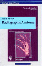
Pocket Atlas of Radiographic Anatomy
Normal findings in CT and MRI

Torsten B. Moeller, M.D.
Am Caritas-Krankenhaus
Dillingen/Saar
Germany
Emil Reif, M.D.
Am Caritas-Krankenhaus
Dillingen/Saar
Germany
210 Illustrations
Subscribe to:
Posts (Atom)


